
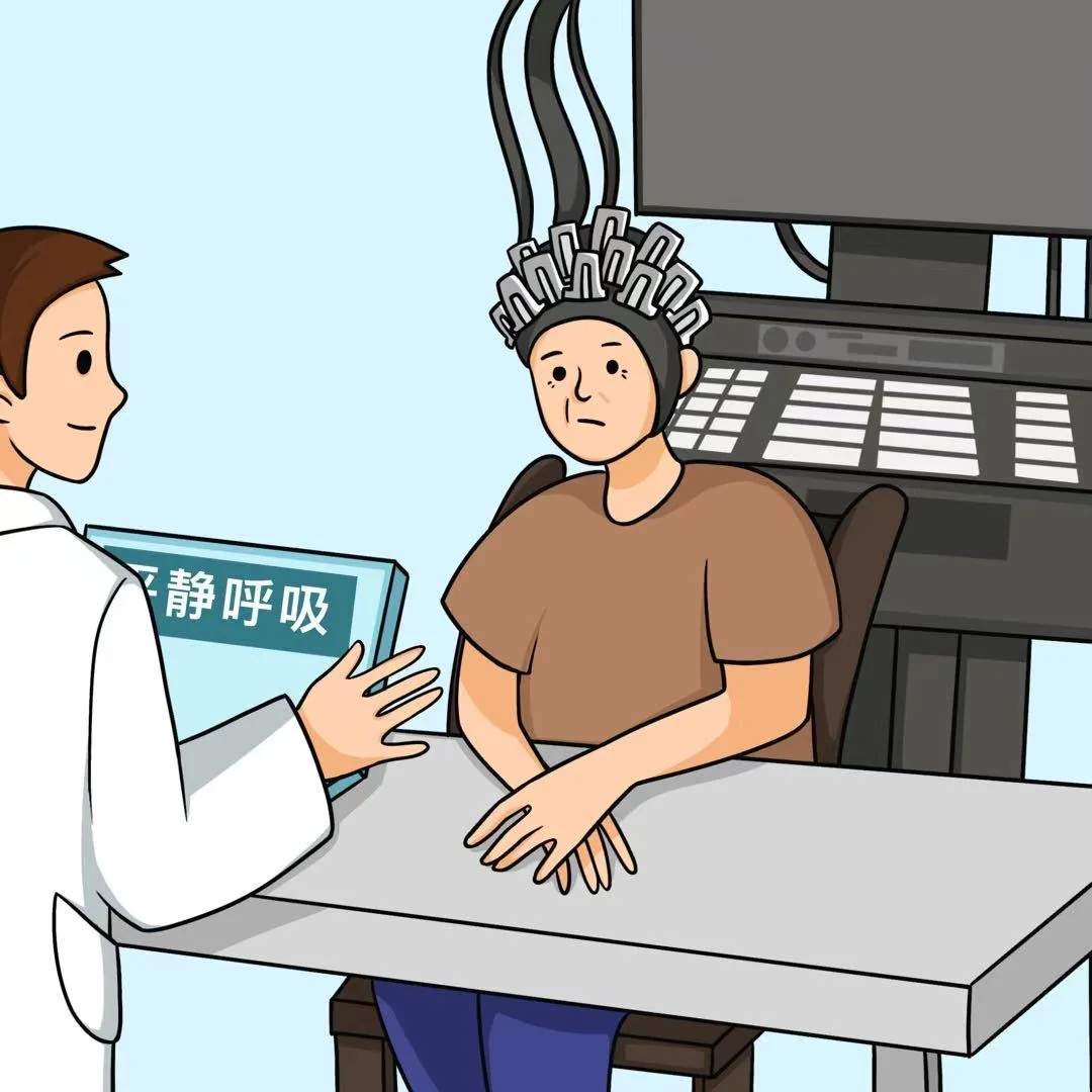
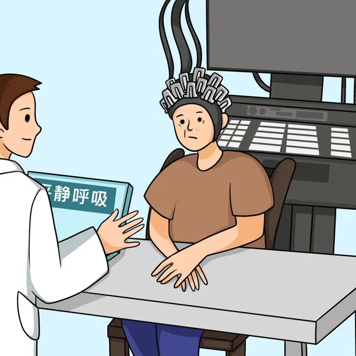
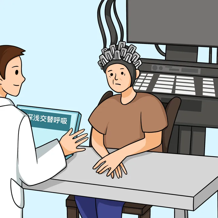
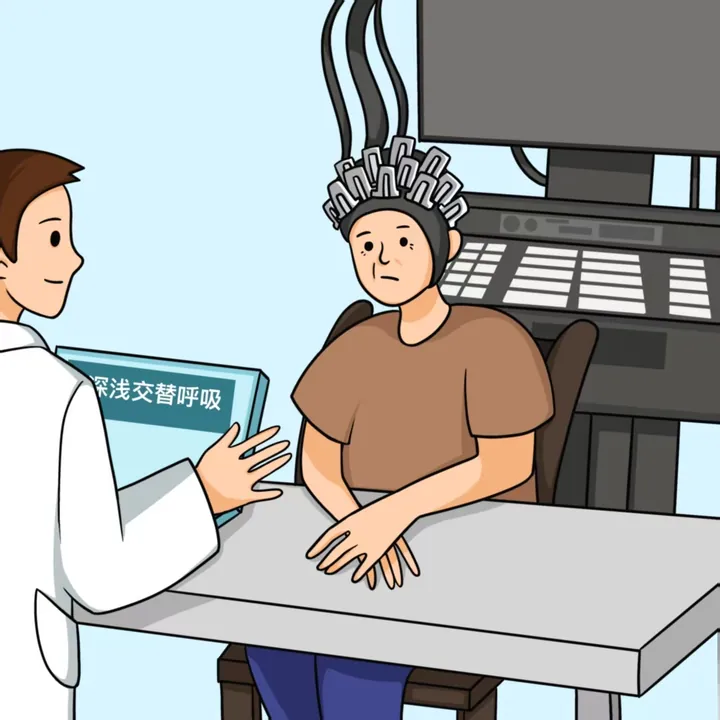
画面呈现脑电实验的典型场景:医生为受试者佩戴电极,屏幕标注“静息态”。这类安静、合作的测量场景,是脑功能研究里最基础的一环。文件名含 breath 字样,但不足以证明画面对应呼吸模式研究;本页按静息态脑电场景归档。
图背后的故事
目的:睡眠剥夺会对健康产生多方面影响,但睡眠与心血管系统功能的关系尚不清楚。研究探讨睡眠剥夺后大脑与心音的变化。方法:招募 90 名健康成年男性,进行 36 小时睡眠剥夺,每 2 小时测量心音、血氧和心率;通过主成分分析对心音数据降维,并在睡眠剥夺前后用功能磁共振成像测量低频振幅与局部一致性指标,再用相关及回归分析考察脑功能与心音变化之间的联系。结果:36 小时内心率和血氧未见异常值,心音强度则出现明显升降波动;多个脑区的功能指标发生变化。回归分析提示,左侧距状皮层的变化可解释收缩期、第一及第二心音强度的变化。结论:急性睡眠剥夺影响心脑轴及特定脑区,睡眠剥夺期间左侧距状皮层的变化参与心脏收缩调节。
论文摘要(中文译文 · 原文)
中文译文 目的 睡眠剥夺(SD)因其多方面的不良影响已成为现代社会面临的健康问题,但睡眠与心血管系统功能之间的关系仍不明确。本研究探讨睡眠剥夺后大脑与心音的变化。方法 招募90名健康成年男性,接受36小时睡眠剥夺,每2小时测量心音、血氧与心率等;采用主成分分析对心音数据降维,在睡眠剥夺前后以功能磁共振成像(fMRI)测量 ALFF 与 ReHo 指标,并用相关与回归分析揭示睡眠剥夺所致 fMRI 与心音变化之间的关系。结果 36小时睡眠剥夺期间,心率与血氧无异常值,而心音强度出现显著的升降波动。双侧距状沟(Calcarine)、左侧楔前叶(Precuneus_L)、右侧颞上回(Temporal_Sup_R)与左侧辅助运动区(Supp_Motor_Area_L)的 ALFF 升高;右侧额上回内侧(Frontal_Sup_Medial_R)、右侧背外侧额上回(Frontal_Sup_R)与左侧内侧额回(Frontal_Mid_L)的 ALFF 降低。回归分析发现,收缩期、S1 与 S2 时相的心音强度可由左侧距状沟(Calcarine_L)的变化解释。结论 急性睡眠剥夺影响心脑轴及特定脑区;睡眠剥夺期间左侧距状沟的变化参与心脏收缩的调节。
Aims Sleep deprivation (SD) has become a health problem in modern society due to its adverse effects on different aspects. However, the relationship between sleep and cardiovascular system function remains unclear. Here we explored the changes occurring in the brain and the heart sounds after SD. Methods Ninety healthy adult men were recruited and subjected to 36 h of Sleep Deprivation (SD). They participated in a number of tests, including measurements of the heart sound, blood oxygen, and heart rate every 2 h. By using of principal component analysis to reduced the dimensionality of heart sound data. While the ALFF and ReHo indexes were measured via fMRI before and after SD. Correlation and regression analyses were used to reveal the relationship between fMRI and heart sound changes due to SD. Results In this study, there were no abnormal values in the heart rate and blood oxygen during 36 h of SD, whereas the intensity of heart sounds fluctuated significantly increased and decreased. The ALFF was increased in bilateral pericalcarine(Calcarine), left anterior cuneus, (Precuneus_L), right superior temporal gyrus(Temporal_Sup_R), left supplementary motor area (Supp_Motor_Area_L); However, it was reduced in the right medial superior frontal gyrus (Frontal_Sup_Medial_R), right dorsolateral superior frontal gyrus (Frontal_Sup_R) and left medial frontal gyrus (Frontal_Mid_L). The regression analysis uncovered that the intensity of the heart sound in the systole, s1, and s2 phase could be explained by Calcarine_L changes. Conclusion Acute sleep deprivation affects cardiac-brain axis and the specific brain regions. Calcarine_L changes during sleep deprivation are involved in regulating heart contractions.