
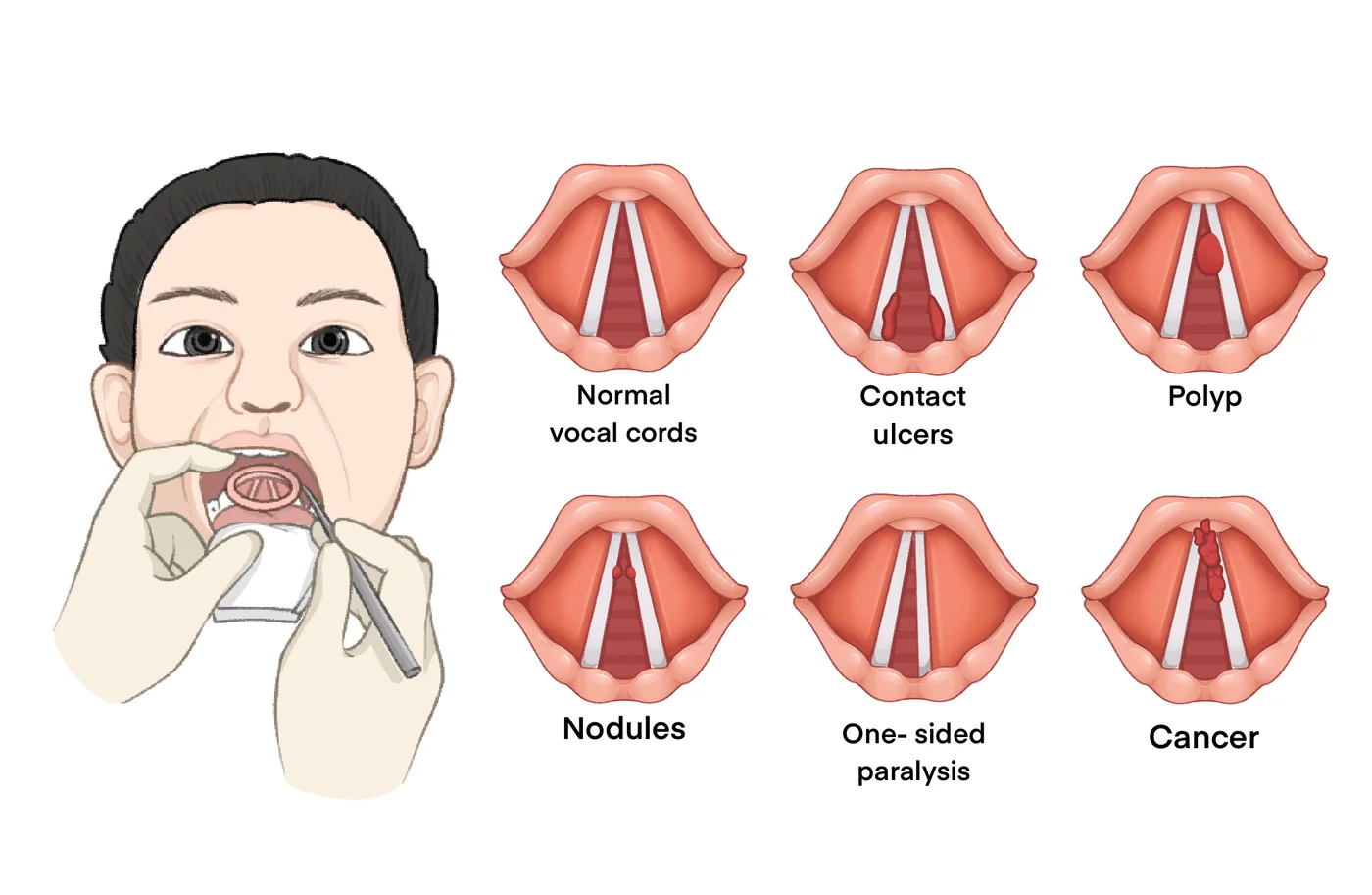
Two parts: a comparison chart of normal vocal folds against polyps, nodules and unilateral paralysis (labels in English), and a silent ~5-second phonation animation. Related reviews discuss acoustic features of organic lesions and AI in voice research; this site does not offer diagnosis.
The Story Behind the Image
Paper abstract (original)
Abstract Objective Voice disorders resulting from organic vocal cord lesions, whether benign or malignant, often lack reliable non-invasive diagnostic tools, which can lead to delays in treatment. This study aims to identify distinctive acoustic biomarkers and develop machine learning models for accurate classification of these lesions and prediction of malignancy. We investigated the acoustic characteristics of voice production in patients with organic vocal cord lesions, comparing benign and malignant cases, and evaluated the diagnostic potential of machine learning models in distinguishing between healthy and pathological voices. Methods A total of 157 participants were enrolled, including 127 patients with organic vocal cord lesions (109 benign, 18 malignant) and 30 healthy controls. Acoustic analysis was performed on vowel sounds, assessing vocal fold vibration parameters. Machine learning models (eXtreme Gradient Boosting (XGBoost), Light Gradient-Boosting Machine (LightGBM)) were trained to classify lesion types and predict malignancy. Receiver operating characteristic analysis identified key diagnostic parameters. Results Comparative analysis revealed 63 statistically significant differences in acoustic parameters between healthy and lesion-affected groups, with skewness and kurtosis being particularly discriminative. Six key parameters (/u/skew, /i/skew, /o/kurt, /o/shapefactor, /o/impulsefactor, and /a/peak2valley) demonstrated high diagnostic value in distinguishing benign from malignant lesions. The XGBoost model achieved the best performance in classifying vocal cord lesions (area under the curve (AUC) = 0.735), while LightGBM excelled in malignancy prediction (AUC = 0.924). Age and specific acoustic parameters were significant predictors in the models. Conclusion The integration of acoustic analysis with machine learning significantly enhances the diagnostic accuracy for vocal cord lesions, particularly in differentiating between benign and malignant cases. These findings underscore the potential of artificial intelligence (AI)-assisted voice analysis as a non-invasive tool for early detection and clinical decision-making. Further validation in larger cohorts is necessary to refine predictive algorithms for broader clinical application. Keywords: Vocal cord lesions, acoustic analysis, objective evaluation, machine learning models, voice disorder diagnostics